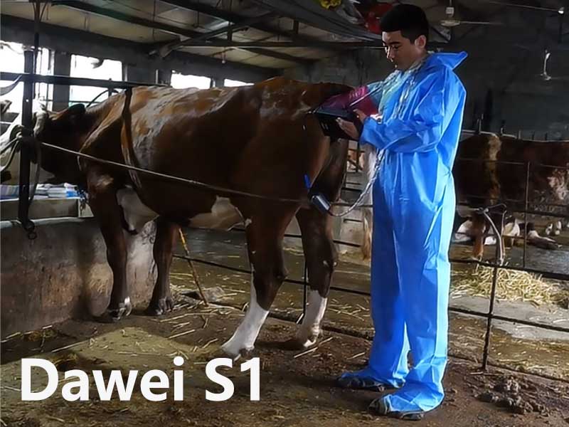
Equine Reproductive Ultrasound
Ultrasound examination is an important and commonly used tool for evaluating the reproductive system of horses. It provides a non-invasive method to examine the reproductive organs of mares and stallions. This article will detail the steps and key observation points for equine reproductive ultrasound examination.
Equine Reproductive Ultrasound Examination
Uterus and Ovaries Ultrasound Examination
Start from the left side of the abdomen, placing the ultrasound probe in the paralumbar fossa area, and move forward towards the pelvic area.
Uterus
- Body: First, locate the uterine body. On the ultrasound image, the uterine body typically appears as a medium echogenic structure, which may contain fluid or other echogenic materials, depending on the mare’s reproductive cycle.
- Horns: From the uterine body, extend to the uterine horns on both sides. The shape and size of the uterine horns will vary based on the mare’s reproductive cycle and health status.
- Left Ovary: By moving the probe upward and slightly towards the midline, the left ovary can be located. Ovaries usually appear as low to anechoic structures, potentially containing multiple follicles or corpora lutea.
- Right Ovary: Repeat the same steps to locate the right ovary. Pay attention to the symmetry between the two ovaries and any internal structural changes.
Ovaries
Pregnancy Examination
Early pregnancy ultrasound should particularly focus on the presence of an embryonic vesicle within the uterus. The embryonic vesicle can typically be detected via ultrasound 14-16 days after conception. As the pregnancy progresses, the structures of the fetus and placenta will become more defined.
Key Observation Points
- Embryonic Vesicle: In the early stages of pregnancy, the embryonic vesicle appears as a round or oval anechoic structure.
- Fetal Heartbeat: Around 20 days of gestation, the fetal heartbeat can be detected via ultrasound.
- Placenta: In mid to late pregnancy, the placenta appears as a highly echogenic structure, and its thickness and uniformity should be checked.
Stallion Reproductive Ultrasound Examination
Testes and Epididymis Examination
The reproductive ultrasound examination of stallions primarily focuses on evaluating the testes and epididymis.
Testes
- Position: Place the ultrasound probe in the scrotal area. The testes should appear as symmetrical, oval structures.
- Internal Structure: The internal structure of the testes should be uniformly medium echogenic. Look for any abnormal echogenic changes, such as cysts or calcifications.
- Head and Tail: The head and tail of the epididymis are located at the front and rear ends of the testes, respectively. The epididymis should appear as a hypoechoic elongated structure.
- Vas Deferens: The tail of the epididymis extends into the vas deferens, which should appear as an anechoic slender tubular structure.
Epididymis
Conclusion
Ultrasound examination holds significant value in evaluating the reproductive system of horses. Detailed examination steps and key observation points enable effective assessment of equine reproductive health. Regular ultrasound examinations help in the timely detection and management of potential issues in the reproductive system, thus enhancing breeding success rates.

