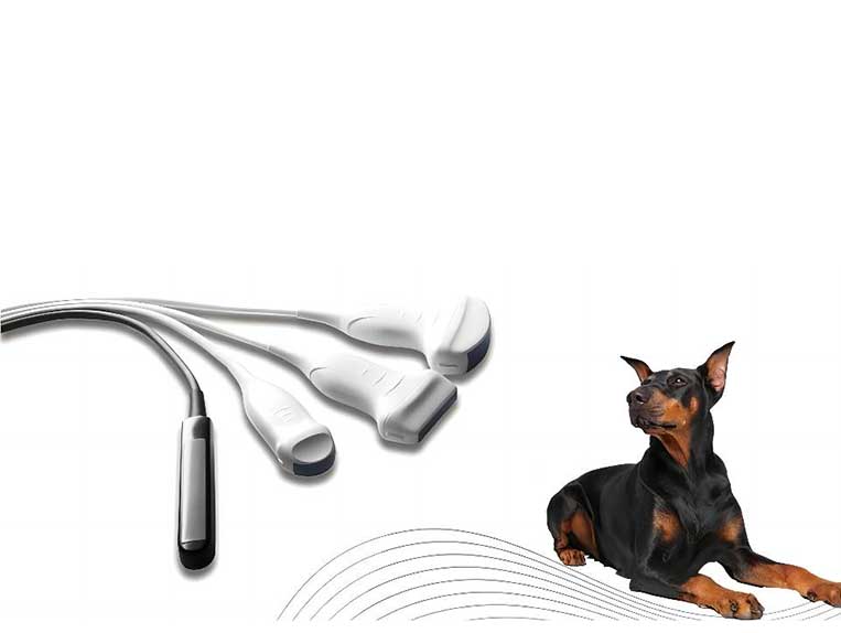
How does the probe frequency of a veterinary ultrasound machine affect ultrasound imaging?
The name or marking of the probe on a veterinary ultrasound system has a number followed by “MHz” and this number (or range of numbers) refers to the frequency of the sound waves produced by that particular probe. As the sound waves travel through the tissue, some of them are absorbed or attenuated, while others are reflected back to the probe to produce an image. Higher frequency sound waves are more affected by attenuation, but because of their shorter wavelength, they are also more accurate in distinguishing between two neighboring structures. In contrast, lower frequency sound waves are less susceptible to absorption but may not be able to discriminate between smaller structures due to their longer wavelength.
What does this mean for you? Higher frequency probes produce higher resolution images but do not penetrate. They are used to image small, superficial structures at shallow depths and at high resolution. Imaging at deeper depths requires powerful low-frequency probes, although the images produced may not have the fine detail of images produced at higher frequencies. Therefore, when operating an ultrasound system, it is prudent to select a transducer with the appropriate frequency for the chosen application.
Most modern probes today are broadband, which means they can operate over a range of frequencies. A good rule of thumb is to scan at the highest possible frequency for the penetration you need to achieve so that you can optimize the resolution of the image regardless of depth. Increasing the frequency is a good way to increase the resolution of the image, and if you want to reach deeper structures, you can decrease the frequency.

