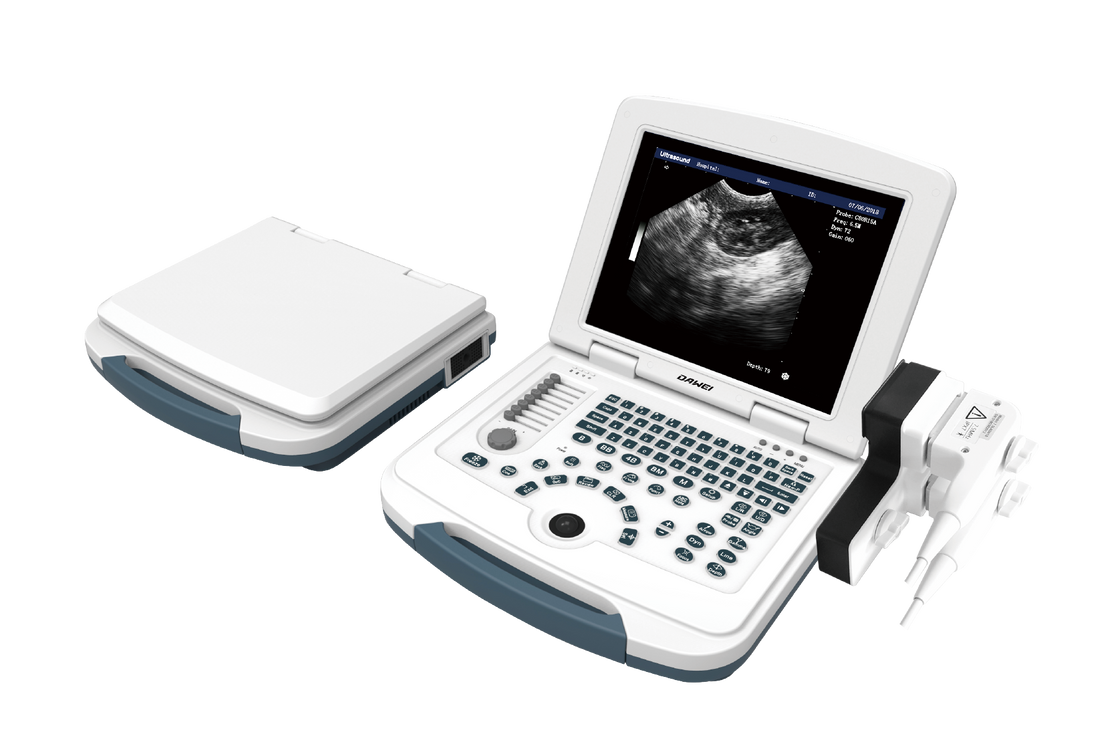
Common Uses of the MU10 Veterinary Sonography Machine
1. Bladder assessment and bladder puncture
Bladder puncture can obtain sterile urine samples, while avoiding complications caused by manual compression and urine extraction, catheterization, or urine contamination problems obtained by natural urination. Ultrasound-guided puncture can reduce the risk of needle puncture of surrounding tissues (such as the colon, posterior vena cava, or descending aorta) and can perform a simple bladder assessment. Although some bladder pathological changes are very mild (such as mild cystitis) and are easily overlooked, other disease changes are obvious. Bladder stones may have different shapes and sizes, but when the stones are large enough, they may produce a high echo interface with posterior acoustic shadowing. For animals with hematuria, blood clots of different shapes may float in the urine or adhere to the bladder wall. Bladder tumors may have different shapes and echogenicity, but tumors generally originate from the bladder wall, showing irregular edges and protruding into the bladder cavity. If a tumor is found in the bladder, bladder puncture is not recommended to prevent tumor cells from spreading along the soft tissue of the needle track.
2. Diagnosis of pleural and peritoneal effusions
Animals with pleural effusions have a variety of clinical signs, many of which are nonspecific, as pleural effusions are complications of a variety of disease processes. Ultrasound is very useful in identifying and sampling pleural effusions in dogs and cats, and is more sensitive than X-rays, and can diagnose even small effusions. The main cavities that require diagnosis of effusions include the abdomen (peritoneal cavity), chest (thoracic cavity), and pericardial cavity.
Abdominal ultrasound uses a lateral or supine position for scanning, but when there is only a small amount of peritoneal effusion, the lateral scan is more likely to detect effusions on the gravity side. When scanning for pleural effusions, the animal needs to be in a standing or prone position, starting from the costochondral junction between the 8th and 9th intercostal spaces, which is also the recommended area for thoracentesis. When evaluating pericardial effusions, it is necessary to scan at the costochondral junction between the 3rd and 4th intercostal spaces on the right side, which provides the best acoustic window for the heart.
Third cavity effusions usually appear anechoic on ultrasound, but this may vary depending on the type of effusion. The echogenicity and cytological grading of effusions are affected by their cellular and protein components, and their echogenicity increases in the following order: simple transudate (anechoic) to modified transudate (multiple different echogenic changes) and exudate (higher echogenicity); however, the echogenicity of intracavitary effusions can also vary, so fluid analysis is required to diagnose their exact nature.
3. Auxiliary diagnosis of gastrointestinal diseases
The most common gastrointestinal symptoms in dogs are anorexia, vomiting, diarrhea, and weight loss. Ultrasound examination focuses on the thickness, stratification, and motility of the stomach, pylorus, gastric wall, and intestinal wall. However, due to the presence of gas in the stomach, the effect of ultrasound examination may not be ideal.
For dogs with vomiting, gastric dilatation with gastric effusions, such as hypercalcemia or pancreatitis-induced gastric retardation, or pyloric outflow obstruction, should be excluded. Ultrasound examination may also reveal foreign bodies, tumors, and strictures in the gastrointestinal tract. Another possible finding is thickening of the stomach wall due to gastritis or tumors. The distribution of lesions, the severity of thickening, and the affected gastric wall layers can assist in differential diagnosis. However, diagnostic tests such as ultrasound-guided biopsy or endoscopy are often required to confirm the diagnosis. In dogs with chronic diarrhea and weight loss, gastrointestinal ultrasound examination may help exclude diffuse or focal intestinal wall thickening caused by inflammatory or neoplastic diseases.
4. Identification of liver and spleen masses
Nodules or masses in the liver and spleen can be primary or metastatic; can be benign or malignant; and may appear as a single mass, multiple masses, or diffuse infiltration. The detection of soft tissue nodules is affected by the sonographer's skill level, ultrasound probe resolution, and the echogenicity of the surrounding parenchyma. Common malignant liver and spleen tumors include hepatocellular carcinoma, angiosarcoma, lymphoma, and histiocytic sarcoma. Common benign liver and spleen lesions include hyperplasia, myelolipoma, hematoma, lymphoid hyperplasia, and extramedullary hematopoiesis. In liver lesions, large liver masses with peritoneal effusions are usually more suggestive of malignancy than benign hyperplasia. In spleen lesions, multiple nodules with a diameter of 1-2 cm, multiple nodules with a target sign (hyperechoic center and hypoechoic outer ring), and associated peritoneal effusions are suggestive of malignancy. Liver and spleen masses may have different characteristics, including well-defined or poorly defined margins, mixed echoes, intracavitary fluid, and the presence of mineralization. The splenic vein should also be evaluated to exclude thrombotic disease, such as in cases of splenic torsion, protein-losing nephropathy, etc.
5. Evaluation of gallbladder mucocele
The gallbladder is located to the right of the midline, between the right lateral lobe and the quadrate lobe of the liver. The normal gallbladder contains anechoic bile and has a thin gallbladder wall (cat: 1mm; dog <2mm). Cats may occasionally find a lobulated gallbladder, which is normal. Bile is normally anechoic, but sludge-like material of varying echogenicity may be seen in the gallbladder, either as suspended particles or as sediment on the gravity side. Echoic gallbladder contents may be an incidental finding and unrelated to biliary disease.
Hypoechoic mucus deposits in a distended or full gallbladder may be classified as gallbladder mucocele, but may also be bile concentrate, myxoid hyperplasia, cystic hyperplasia, and other related conditions. Gallbladder mucocele is common in older small dogs and is rare in cats. The development of gallbladder mucocele is continual, starting with echogenic bile, progressing to a stellate appearance, and finally to the kiwi fruit sign. Although the exact etiology of gallbladder mucocele is controversial, its clinical significance is significant, resulting in gallbladder rupture, bile peritonitis, and the need for emergency surgery.
Elective cholecystectomy (before gallbladder rupture or biliary obstruction) is associated with lower mortality than non-elective cholecystectomy (after gallbladder rupture or biliary obstruction). Suspended mucus (not gravity-side sediments) and/or hyperechoic bile are potential indications for cholecystectomy.
Although mature gallbladder myxomatosis will demonstrate gallbladder enlargement and thin gallbladder wall, there are many etiologies (eg, cholecystitis, gallbladder wall edema, gallstones, gallbladder mucosal hyperplasia, and tumors [rarely]) that can lead to thickened gallbladder wall (>3-3.5 mm). Ultrasound-guided gallbladder puncture and bile aspiration can be performed for bile culture and/or gallbladder decompression. However, this procedure carries an increased risk of bile leak (secondary bile peritonitis) in animals with a dilated gallbladder or gallbladder wall disease.
