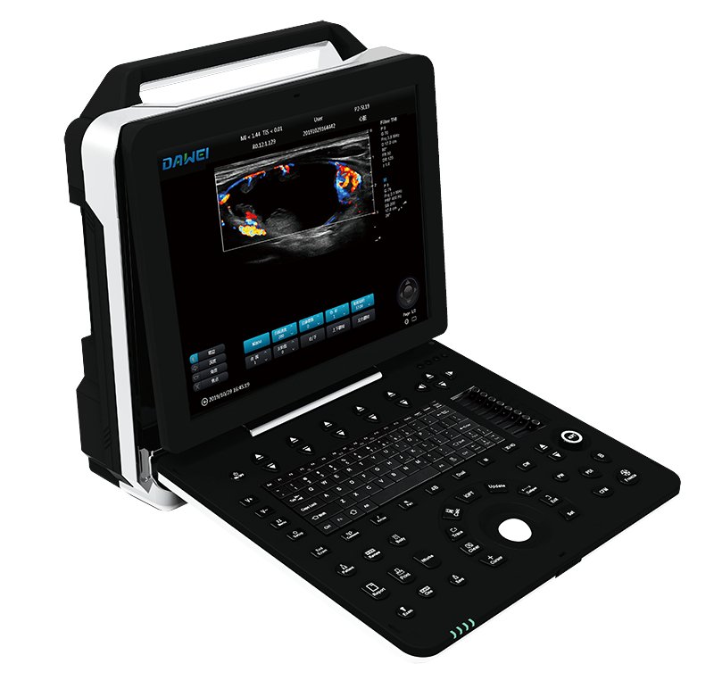

Color Doppler Veterinary Ultrasound Scanner P8-VET
Color Doppler Veterinary Ultrasound Scanner P8-VET
Couldn't load pickup availability
|
1. Product Name: Full Digital Color Doppler Veterinary Ultrasound Scanner |
|
1.1. Structure Style: Portable |
|
2. Application 2.1. Applicable for animals’ examination and diagnosis on digestive system, reproductive system, urinary system for animal hospitals and scientific institutions. |
|
3. System Technical Specifications and Summary 3.1. Full digital color doppler ultrasound diagnostic system |
|
3.2. Digital Beam Enhancer |
|
3.3. Multiple Beam Synthesis |
|
3.4. Full-Digital 2D Gray Scale Imaging |
|
3.5. Tissue Harmonic Imaging (THI) |
|
3.6. B/C Real-time Two Synchronous Imaging |
|
3.7. M Mode Imaging |
|
3.8. Anatomic M Mode Imaging (Sampling line≥3) |
|
3.9. Color Doppler Imaging (CFM, PDI, DPDI) |
|
3.10. Spectral Doppler Imaging (PW, HPRF PW, CW) |
|
3.11. Tissue Doppler Imaging (TVI, M-mode, Spectral Imaging, etc.) |
|
3.12. Four-dimensional Supersonic Image Formation |
|
3.13.★Contrast Tuned Imaging |
|
3.14.★PView Wide Field Imaging |
|
3.15. Space compound imaging (application to Abdominal, GYN, vessel, superficial small organs, can be dual image contrast display at same time) |
|
3.16. Frequency & Focus Compound |
|
3.17. Extended field-of-view (EFOV) |
|
3.18. Real-time dual image contrast display |
|
3.19. B/C/D Real-time three synchronous imaging |
|
3.20. Speckle Reduce Imaging (SRI) |
|
3.21. Elastography |
|
4.★Operation Interface: support 10 different languages |
|
5. System Specification 5.1. Monitor: 15 inch high resolution medical LCD monitor |
|
5.2.★Probe connectors: ≥2 active, support all probes |
|
5.3. 2D Imaging Mode 1) Full Digital Beamformers 2) Digital full dynamic focusing, digital variable aperture and dynamic trace change, A/D≥15 bit 3) Receiving mode: transmitting and receiving channel ≥1024, multi-signal parallel processing 4) Line density: ≥512, visible and adjustable 5) Under B mode, focus number≥10 level, focus position continuously adjustable 6) TGC: 8 segment adjustable 7) Gain: B / M / D separately adjustable, ≥100dB 8) ★Dynamic range ≥180dB 9) ★Max scanning depth ≥360mm 10)Grey scale: 0-67, visible and adjustable 11)Sound power: 1%-100% 12)Linear array probe two-dimensional independent deflection 13)ZoomPIP (1.5 / 2.0 / 2.5 / 3.0 / 3.5 / 4.0 / 4.5 / 5.0 / 10 times) |
|
5.4. Color Doppler Imaging Mode 1) Imaging: including velocity, velocity variance, energy, direction energy display, etc 2) Display mode: B/C, B/C/M, B/POWER, B/C/PW 3) Line density ≥3 level 4) Color hiding technology: without back to B mode can hide C mode, only color speed ruler display 5) Blood flow graph, color flow profile to measure intravascular velocity |
|
5.5. Spectral Doppler Imaging 1) Display: full screen, duplex / triplex (PW only) 2) Gain ≥100dB 3) Velocity ≥4 level adjustable 4) Max scanning speed: PWD: Positive or negative blood flow velocity≥7.6m/s; CWD: blood flow velocity ≥20.0m/s, min ≤5 mm/s (Nonnoise signal); 5) Zero shift adjustable ≥8 level 6) Display mode: B, PW, B/PW, B/C/PW, B/CW, B/C/CW, etc 7) Automatic Spectrum Measurement, Manual Measurement
8) Display control: reverse, zero shift, B refresh, D extension, B/D extension, etc 9) Smart Doppler technology: freely switch between real-time B+CFM mode and real-time PW mode |
|
5.6. Four-dimensional Supersonic Image Formation 1) 4D imaging mode: Surface mode, maximum mode, minimum mode, perspective mode, etc 2) Display mode: B,2B,4B 3) Clipping function; 4) Transparency:130-490;19 level adjustable 5) Threshold value:0-130 6) Smoothness≥4 7) 4D format stores images, movies, volume data |
|
5.7. ★PView wide field imaging(Optional) 1) High-resolution display, length can up to 50cm 2) Imaging support forward erasure, no need to re-imaging 3) With two dimensional wide view imaging mode and color wide view imaging mode |
|
5.8. Probe interface ≥2, fully activated. The volume probe is plug and play. |
|
5.9. Probe: wideband with frequency conversion, independent frequency conversion under B and CFM 1) Fundamental Frequency Conversion≥3 level; 2)1. Convex probe Fundamental frequency: 2.0MHz/2.3MHz/2.5MHz/3.0MHz/3.5MHz/4.0MHz/4.5MHz/5.0MHz/5.5MHz,nine levels frequency conversion(depth:50-360mm) Harmonic Frequency:10.0MHz/14.0MHz/18.0MHz,three levels frequency conversion
1.7MHz/1.9MHz/2.0MHz/2.5MHz/3.0MHz/3.4MHz/3.8MHz/4.2MHz/5.5MHz, nine levels frequency conversion(depth:50-360mm) Harmonic Frequency:3.0MHz/4.0MHz/5.0MHz,three levels frequency conversion 3.0MHz/3.5MHz/4.0MHz/5.0MHz/5.4MHz/6.0MHz/7.0MHz/8.0MHz,eight levels frequency conversion(depth:50-360mm) 8.Micro-convex probe R11/R15: 3.0MHz/3.5MHz/4.0MHz/5.0MHz/5.4MHz/6.0MHz/7.0MHz/8.0MHz,eight levels frequency conversion(depth:30-160mm) Fundamental frequency: 3.0MHz/3.5MHz/4.0MHz/5.0MHz/5.4MHz/6.0MHz/7.0MHz/8.0MHz/10.0MHz,nine levels frequency conversion(depth:40-120mm)
Remark:The above probes have harmonic frequencies Remark:Optional according to customer needs:Convex probe, linear probe, micro convex probe, phased array probe, volume probe, etc |
|
6. Measurement / Analysis and Reports 6.1. General measurement: distance, area, ellipse, cross line, angle, distance ratio, volume, volume (ellipse), area ratio, diameter, joint angle. |
|
6.2. Specialized measurement: 1) Cardiac:LV、MPAD、RVEDd、RVEDs、LVM、LAVol、RV/LV、LVSimps、LA/AO、MV、LVMassA/L、QP/QS. 2) ★OBC:Canine:GSD, CRL, HD, BD; Feline: BD, HD; Swine: HLA, SLA; Bovine: CRL, BTD, BUD;Ovine: SCRL, BPD;Equine: GSD(H), ESD (V) |
|
7. Peripherals Section 7.1. Configured ultrasonic graphic work station, the work station software equipped with registration certificate, supports black and white digits, simulation B/W, digit color, simulation color, text and video printer, and foot switch 7.2. Support Internet connection 7.3. Support DICOM 3.0, report of DICOM Obstetrics and Gynecology / Heart / Blood Vesse 7.4. Video/Audio Input Output 7.5. Main unit comes with USB port 7.6. Support ultrasonic system directly sent clinical images and reports through Internet. |
|
8. Cine Loop and Original Data Processing 8.1. Cine Loop ≥ 3061 frames, support manual and auto operation 8.2. Digitalization Hard Drive Capacity ≥ 256G, support permanent store static, dynamic images, can be consulted, transferred, deleted freely 8.3. Multiple Image Export Modes: Dynamic Image and Static Image exported in PC format, Images can be seen on regular PC devices, no need of specific software. Can process diagnosis, export and backup image data at the same time without affecting the inspection operation 8.4. Professional probe holder ≥2 (coupling holder not included),each holder can be charged left and right |