Skip to product information
1 of 2

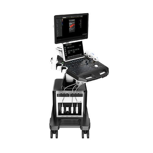
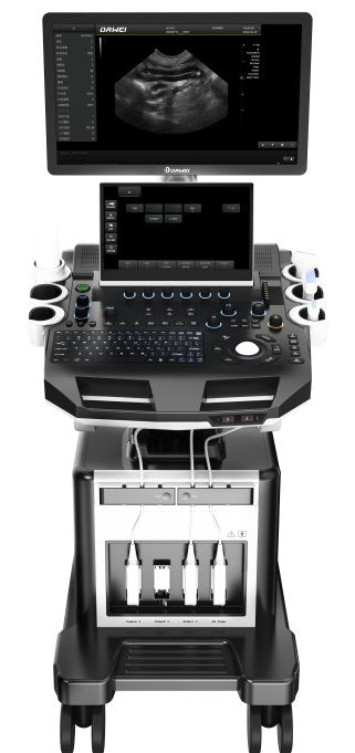
DWanimal T3-VET Veterinary Color Doppler Ultrasound

DWanimal T3-VET Veterinary Color Doppler Ultrasound

Regular price $59,154.00 USD
Regular price $9,999.00 USD Sale price $59,154.00 USD
Sale Sold out
Select your probe(s)

1.Product name: Veterinary Color Doppler Ultrasound Diagnostic System

1.1 Structure type: Trolley with two screens

2. Application:

2.1 Applicable for animals’ ultrasonic examinations in animal hospital,zoo,breeding base and scientific research institution 

3.Technical Specifications and Summary:

3.1 Operation System:Windows10

3.2 Pulsed wave Doppler

3.3 Directional Energy Doppler

3.4 Real-time Triplex

3.5★Spatial Compound Imaging

3.6 Tissue Harmonic Imaging (THI)

3.7 2B/4B Imaging Mode

3.8 System language: Chinese, English, French, Russian and Spanish

3.9 Display size: ≥ 21.5 inches

3.10 Integrated clipboard: display the saved images at the bottom of the screen, can be directly saved or deleted

3.1 System with on-site upgrade function

3.12 Preset conditions: for different inspections, preset inspection conditions to optimize the image, reduce the operation of the adjustment, commonly required external adjustment and combination adjustment

3.13 Support real-time 3D imaging function

3.14★Probe connector ≥ 4

3.15★Trapezoidal imaging function

3.16 One key intelligent optimization function

4. Probes :

Linear Probe:Frequency 6.0MHz/7.5MHz/8.5MHz/10.0MHz/12.0MHz/H10.0MHz, six-segment frequency conversion (detecting depth 20-128mm)

Micro-convex(R11):Frequency 4.5MHz/5.0MHz/6.0MHz/6.5MHz/7.0MHz/9.0MHz/H8.0Mhz, seven-segment frequency conversion (detecting depth 30-111mm)

Micro-convex(R15):Frequency 4.5MHz/5.0MHz/6.0MHz/6.5MHz/7.0MHz/9.0MHz/H8.0Mhz,seven-segment frequency conversion (detecting depth 30-111mm)

Convex Probe:Frequency 2.0MHz/2.5MHz/3.0MHz/3.5MHz/4.0MHz/5.5MHz/H4.0MHz/H5.0MHz,eight-segment frequency conversion (detecting depth 30-255mm)

Rectal Probe:Frequency 4.0MHz/6.5MHz/9.0MHz/H8.0Mhz,four-segment frequency conversion (detecting depth 20-110mm)

High frequency linear Probe (L25): Frequency 6.0MHz/7.5MHz/8.5MHz/10MHz/12MHz/H10MHz six-segment frequency conversion(detecting depth 2-110mm)

Phased array Probe:Frequency 2.0MHz/2.5MHz/3.0MHz/3.5MHz/4.0MHz/5.0MHz/H3.0MHz/H4.0MHz eight-segment frequency conversion (detecting depth 10-244mm)

Volume Probe:Frequency 2.0MHz/3.0MHz/4.0MHz/4.5MHz/5.5MHz/6.0MHz/H5.0MHz seven-segment frequency conversion (detecting depth 30-237mm)

Backfat Probe:Frequency 2.0MHz/3.5MHz/4.0MHz/5.0MHz/H4.0MHz/H5.0MHz six-segment frequency conversion (detecting depth 30-237mm)

5.2 D Imaging Mode:

5.1 Gain: 0-100, Step 1 adjustable

5.2 TGC: 8 segment adjustable

5.3 Dynamic: 20-280, 20 levels adjustable

5.4 Pseudo color: 0-11, adjustable

5.5 Sound power: 5%-100%, step 5% adjustable

5.6 Body mark: ≥17 kinds optional

5.7 Maximum focus: ≥6, which can be moved throughout the whole process

5.8 Grey scale map: 0-7, 7 levels adjustable

5.9 Filter: 0-4

5.10 Scanning range:  50%-100%

5.11 Frame correlation: 0-4, 4 level adjustable

5.12 The screen has real-time display of sound power, probe frequency, dynamic range, pseudo color, gray scale and other 14 parameters can be adjusted.

5.13 Line density: low, middle, high, 3 levels adjustable

5.14 Noise reduction: 0-14

6. Color Doppler Imaging Mode

6.1 Color Frame correlation: 0-12, 12 levels adjustable

6.2 Color map: 0-7, 7 levels adjustable

6.3 Color flip: adjustable

6.4 B / C real-time split screen mode

6.5 Base line: 11 levels, adjustable

6.6 Line density: low and high adjustable

6.7 Filter: 0-5 levels adjustable

7. Spectral Doppler Imaging Mode

7.1 Sampling volume angle correction: -80°~80°adjustable

7.2 Sample volume: 0.5mm-20mm adjustable

7.3 Frequency: 2.5MHz and 3.0MHz visible and adjustable (different probe ranges vary)

7.4 Base line: 11 levels, adjustable

7.5 Pseudo color: 0-5

7.6 Parameter display: ≥4 kinds, adjustable

7.7 Speed scale: 32.8-328cm/s (different probes have different ranges)

7.8 Spectrum envelope function: real time automatic spectrum envelope, manual spectrum envelope, and

other. The system automatically analyses and displays various data such as PS, ED, RI, PI, S/D, HR, etc. 7.9 Grey map: 0-7

7.10 Filter: 0-8

7.11 Dynamic range: 10-95db, step 5

7.12 Noise reduction: 0-28

7.13 Sound volume: 0-100

8. 3D imaging mode (optional)

8.1 Fast angle: support for 0°, 90°, 180°, 270° rotation of 3D window images.

8.2 Display layout: support "double", "quad", "single" image display.

8.3 Reconstruction modes: RealSkin, surface, Max, Min, XRax.

8.4 Pseudo-color display: support 0-7 level adjustment.

8.5 Image magnification: support for 5 levels.

8.6 Contrast ratio: 0%-100%.

8.7 Threshold: 0%-100%

8.8 Smoothing: ≥ 3 adjustable gears

8.9 X-axis, Y-axis, Z-axis rotation support adjustable

8.10 Brightness: 0%-100%.

9. 4D imaging mode (optional)


9.1 fast angle: support for 3D window image 0 °, 90 °, 180 °, 270 ° rotation.

9.2 Display layout: support "double", "four", "single" image display.

9.3 Reconstruction modes: RealSkin, surface, Max, Min, XRax.

9.4 Pseudo-color display: support 0-7 level adjustment.

9.5 Image magnification: support for 5 levels.

9.6 Contrast ratio: 0%-100%.

9.7 Threshold: 0%-100%

9.8 smoothness: ≥ 3 adjustable gears

9.9 pseudo-color: ≥ 7 adjustable gears

9.10 X-axis, Y-axis, Z-axis rotation support adjustable.

9.11 line density: support for two levels of adjustment.

10. Measurement and Analysis

10.1 General measurement: distance, area, angle, time, slope, heart rate, speed, acceleration, blood flow path, blood flow spectrum trace, resistance index/pulsation index, etc

10.2 Professional obstetric measurement data package for canine, feline, equine, bovine and ovine

10.3 Measurement line: color and type can be adjusted at will (including the activation color and the completion color)

10.4 Measurement result: display position and font size can be adjusted as needed

10.5 Professional data package: obstetrics, liver and gallbladder, kidney, bladder, heart, stomach and intestines, uterus, adrenal gland, pancreas, blood vessels, etc

11. Graphic and Text Management System:Image format: BMP, DCM, JPG

11.1Host build in hard disk ≥ 256G , start fast and stable

11.2 Movie playback: ≥600 frames

11.3 Internal file information management system: can record pet ID, pet name, owner name and so on, and can be searched and managed by ID, owner name, pet name and so on.

11.4 Report type: ≥6, Abdominal report, cardiac report, obstetric report, small organ report, urinary report, vascular report, picture proof provided

11.5 One key fast report graphic and text management

12. Interface:

4*USB; 1Video; 1 S-Video; 1 DVI; 1 HDMI; 1 RJ-45;

13. Configuration:

13.1 Full Digital Veterinary Color Doppler Ultrasound Diagnostic System

13.2 Probe:Micro-convex R11 probe(standard), Convex probe(optional), Linear probe(optional), Micro-convex R15 probe (optional)

13.3Video printer (optional), laser printer (optional)

View full details