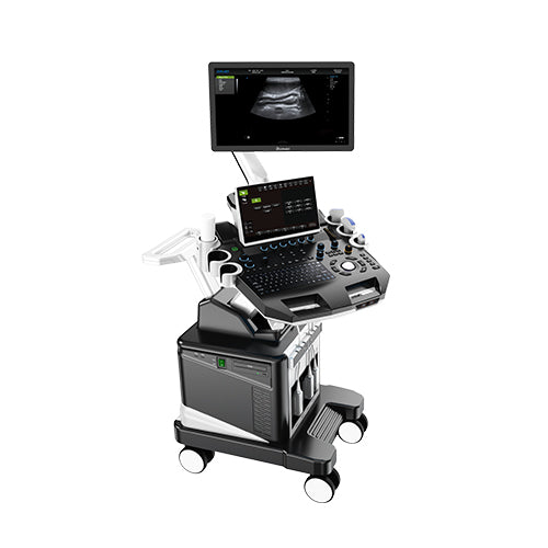
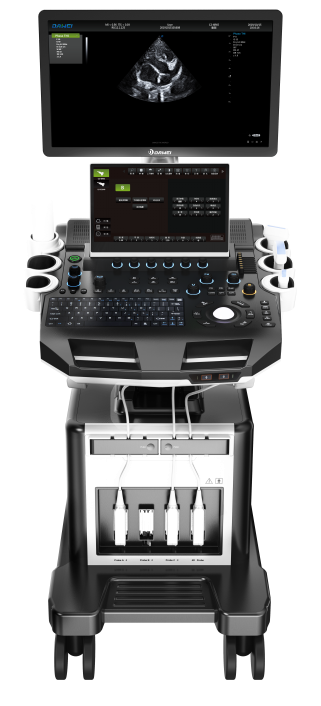
DWanimal T8-VET Veterinary Color Doppler Ultrasound
DWanimal T8-VET Veterinary Color Doppler Ultrasound
Couldn't load pickup availability
|
Product Name:Veterinary Color Doppler Ultrasound Diagnostic Instrument. |
|
|
|
1.1 Structure Style :Dual Screen Cart |
|
22.2. Application: : 2.1 It's applicable for animals’ examination and diagnosis on digestive system, reproductive system, urinary system for animal hospitals and scientific institutions. |
|
|
3. System technical specifications and overview |
|
|
3.1. Full digital color Doppler animal ultrasound diagnostic system mainframe 3.2 Digital beam booster 3.3. Multiple beamforming 3.4. Two-dimensional grayscale mode 3.5. Harmonic Imaging Technology 3.6. B+C dual real-time mode 3.7. M-type mode 3.8. Anatomical M-mode, sampling line ≥ 3 3.9.Doppler imaging (including color, energy, and directional energy Doppler modes) 3.10. Spectral Doppler imaging (including pulsed Doppler, high pulse repetition frequency, continuous wave Doppler) 3.11. Tissue Doppler imaging (including tissue velocity map, M-mode, spectral imaging and other modes) 3.12. 4-D imaging 3.13.★Contrast Imaging Technology 3.14.★With PView wide-view imaging technology 3.15. Spatial composite imaging technology (can be used for abdomen, blood vessels, superficial small organs, and can be displayed on the same screen with double contrast) 3.16. Frequency compound imaging 3.17. Extended Imaging 3.18. Real-time double contrast imaging 3.20. Real-time three-synchronous imaging (two-dimensional, color, spectrum real-time simultaneous imaging) 3.21. Speckle noise suppression technology 3.22 11 kinds of canine specimens, 5 kinds of feline specimens, 2 kinds of porcine specimens, 10 kinds of bovine specimens, 2 kinds of equine specimens, 4 kinds of sheep specimens, 1 kind of rabbit specimens, and 1 kind of rat specimens. |
|
|
4.★Operating interface: The host contains 10 operating language interfaces. |
|
|
5. System technical parameters and requirements 5.1 standard with ≥ 21.5-inch high-resolution color LCD monitor (optional 23.8-inch high-resolution color LCD monitor) 13.3-inch color LCD touch screen, support multi-touch |
|
|
5.2 ★ host built-in probe interface ≥ 4, fully activated, consistent size, interoperable and interoperable |
|
|
5.3. Two-dimensional gray-scale mode 1)Digital acoustic beamformer 2) digital full dynamic focus, digital variable aperture and dynamic variable traces, A/D ≥ 15 bit 3) reception mode: transmitting and receiving channels ≥ 1024, multiples of signal parallel processing 4) scan line: each frame line density ≥ 512 ultrasound lines 5) launch beam focus: launch ≥ 10 segments, focus position with special menu adjustment 6) TGC ≥ 8 segments 7) gain adjustment: B / M / D are independently adjustable, ≥ 100dB 8) ★ dynamic range adjustment: ≥ 300dB 8) ★ maximum display depth ≥ 410mm 9) gray scale: ≥ 67 levels, visually adjustable 10) sound power: 1%-100% 11) two-dimensional independent deflection of the line array probe 12) local amplification (1.5/2.0/2.5/3.0/3.5, 4.0/4.5/5.0/10 times) |
|
|
5.4. Color Doppler imaging 1) imaging mode: including velocity, velocity variance, energy, directional energy display, etc. 2) display mode: B / C, B / C / M, B / POWER, B / C / PW 3) line density ≥ 3 levels 4) Color hiding technology: no need to return to the two-dimensional mode to hide the color, only display the color velocity scale 5) Blood flow distribution map function, color flow profile to measure intravascular flow velocity |
|
|
5.5. Spectral Doppler mode 1) display format: full screen, duplex / triplex (PW only) 2) gain: ≥ 200dB 3) multi-spectrum speed: ≥ 4 adjustable 4) maximum measurement speed: PWD: positive or negative blood flow velocity ≥ 7.6m / s; CWD: blood flow velocity ≥ 20.0m / s, the minimum speed: ≤ 5 mm / s (non-noise signal) 5) zero shift: ≥ 8 levels 6) display mode: B, PW, B/PW, B/C/PW, B/CW, B/C/CW, etc. 7) spectrum automatic measurement, manual measurement 8) display control: inversion, zero shift, B refresh, D extension, B/D extension, etc. 9) Intelligent Doppler technology, can be freely switched between real-time B+CFM mode and real-time PW mode. |
|
|
5.6. Four-dimensional mode. 1) Display mode: single, double and quad. 2) Cropping function. 3) Transparency 1-509: 10 levels adjustable 4) Threshold 0-129 5) Smoothing ≥ 4 6) four-dimensional format to store pictures, movies, body data |
|
|
5.7.★Wide-field imaging as standard (1) Display length up to 50cm at high resolution (2) With two-dimensional wide field, color wide field imaging mode |
|
|
5.8.★Piercing enhancement function (line array probe) 1)The angle and position of the piercing needle can be adjusted 2)With two guidance modes of puncture line and puncture guidance range |
|
|
5.9. Probe interface ≥ 4, fully activated. Volumetric probe plug-and-play. |
|
|
5.10. Probe: broadband frequency conversion probe, two peacekeeping and color independent frequency conversion. 1. Convex probe: 4.0MHz/4.5MHz/6.0MHz/7.5MHz/8.0MHz/10.0MHz/12.0MHz/14.0MHz/20.0MHz nine-segment frequency conversion(detecting depth 20-120mm) 5.0MHz/5.5MHz/6.0MHz/8.0MHz/9.0MHz/10.0MHz/11.0MHz/12.0MHz/15.0MHz, nine-segment frequency conversion(detecting depth 20-120mm) Note: The above probe has harmonic frequency Note: According to customer needs can be optional: convex array probe, linear array probe, micro-convex probe, phased array probe, volume probe, etc. |
|
|
6. Measurement/analysis and reporting. 6.1 Routine measurements: distance, area, ellipse, crosshairs, angle, distance ratio, volume, volume (ellipse), area ratio, diameter, joint angle. 6.2 Specialized special measurements 1) Cardiac measurements: LV, MPAD, RVEDd, RVEDs, LV myocardium, LAVol, RV/LV, LVSimps, LA/AO, MV, LVMassA/L, QP/QS. 2) ★Obstetric measurements: Canine: cephalopelvic diameter, fetal sac, transverse cranial diameter, transverse body cavity diameter; Feline: transverse body cavity diameter, transverse cranial diameter; Porcine: porcine heart, porcine stomach; Bovine: cephalopelvic diameter, transverse trunk diameter, transverse cranial diameter; Sheep: cephalopelvic diameter, biparietal diameter; Equine: fetal sac (H), fetal sac (V) |
|
|
7. Peripherals part. 7.1. Configure a set of ultrasonic graphic workstation, the workstation software needs to have a registration certificate, support digital black and white, analog black and white, digital color, analog color, text and video printer, support foot switch. 7.2 Support network connection 7.3 Support DICOM 3.0 DICOM obstetrics, cardiac, vascular reports 7.4 Video/audio input and output 7.5 Host comes with USB interface 7.6Support ultrasound system to send clinical pictures and reports directly to the computer through the network 7.7 Optional coupler heater |
|
|
8 movie playback and raw data processing. 8.1 Movie playback ≥ 3061 frames, support manual, automatic playback 8.2 Solid state hard disk is 120G Mechanical hard disk is 1T, which can store dynamic and static images permanently, and can access, transfer and delete images at any time 8.3 Multiple export image formats: dynamic images and static images are exported directly in PC format, so that the images can be viewed directly on a normal PC without special software. Export, backup image data at the same time, real-time inspection, does not affect the inspection operation 8.4 Support DVD R / W burning optical drive 8.5 with professional probe placement rack ≥ 5 (excluding coupling agent placement rack) each probe placement rack left and right can be changed |
|
Share


