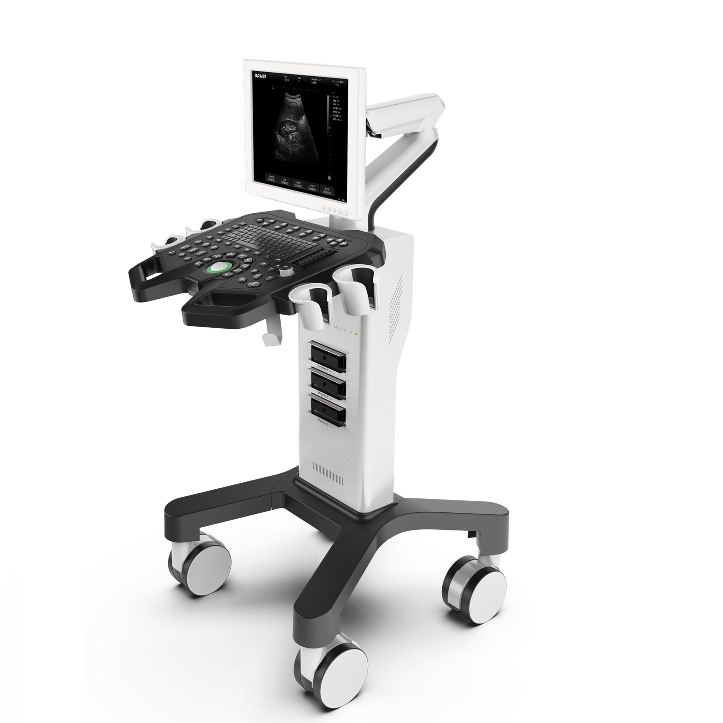
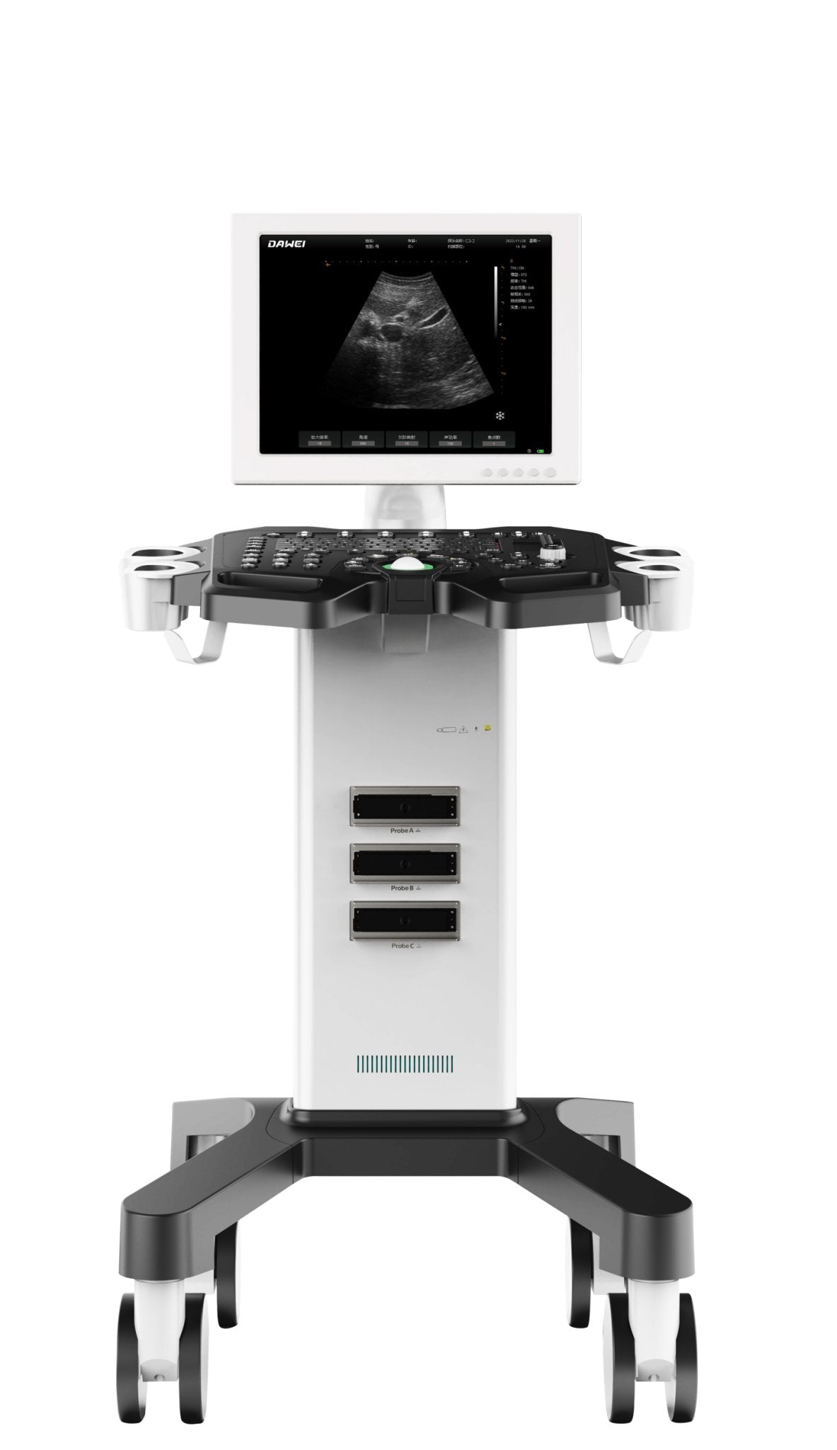
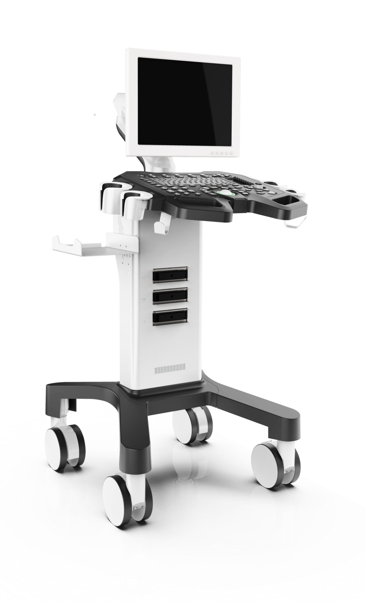
Vetetinary Color Doppler Ultrasound Scanner MT15
Vetetinary Color Doppler Ultrasound Scanner MT15
Spend $489 and get a 20% discount at checkout. 🎁
Couldn't load pickup availability
|
1. Product name: Full digital ultrasound diagnostic system |
|
1.1Structure type: Trolley |
|
2. Description of use and requirements for products 2.1 Ultrasound examination for various needs in pet hospitals, clinics, zoos, breeding/breeding bases, and various research units. |
|
3. Main specifications and system overview: |
|
3.1. Operating system: ARM chip architecture, stable, simple and powerful 3.2. Support three probe sockets, the choice of inspection parts can be made 3.3. Display modes: B, B + B, B + M, M, 4B. 3.4. Display: 15-inch 1024 * 768 resolution high-definition LED LCD screen 3.5. System language: Chinese and English, Spanish, French 3.6. Barebone size: 579.1 * 710.69 * 1220.33mm 3.7. Bare machine weight: 46kg, foam and carton total about 8KG |
|
4. Probes: |
|
4.1. convex probe (C3-2): frequency 2.0MHz/3.0MHz/3.5MHz/5.0MHz/, four frequency bands, the center frequency is 3.5MHz, each frequency band corresponds to the corresponding harmonic frequency (depth adjustment: ≥ 20 levels, adjustment range 50-240mm) 4.2. Linear probe (L3-2): frequency 5.0MHz/7.5MHz/8.0MHz/9.0MHz, four frequency bands, the center frequency is 7.5MHz, each frequency band corresponds to the corresponding harmonic frequency (depth adjustment: ≥ 6 levels, adjustment range 50-100mm) Micro-convex probe (6.5R15Y30C): frequency 5.0MHz/6.5MHz/7.5MHz/8.0MHz, four frequency bands, the center frequency is 6.5MHz, each frequency band corresponds to the corresponding harmonic frequency (depth adjustment: ≥ 8 levels, adjustment range 50-120mm) |
|
5.2D imaging mode |
|
5.1. Gain: 0-100, adjustable in steps of 1 visually 5.2. TGC: 8 segments adjustable 5.3. Dynamic range: 0-120db 5.4. Scan angle: 80-100 5.5. Focus: adjustable focus position, 4 focus points 5.6. Frame correlation: 4-100 5.7. Speckle suppression: 0-50 5.8. Ultrasonic power: 40-100, in steps of 4 5.9. Magnification: 1-16 5.10. Grayscale mapping: 1-19 5.11. Body position markers: ≥ 23 kinds, support body position indicator 5.12. Pseudo-color: 28 kinds 5.13. With lithotripsy positioning function, real-time measurement data 5.14. Support up and down flip, left and right flip, black and white flip 5.15. M super scan speed: 1-63 5.16. Support time setting: date and time, date format, time format, and other settings 5.17. Support automatic freeze setting 5.18. Support automatic sleep time of 6-99 minutes 5.19. Support image storage function: picture saving format: BMP 5.20. Movie playback: 256 Notes support English upper and lower case |
|
6Measurement and analysis functions: |
|
6.1 with conventional measurement functions: distance, perimeter-ellipse, perimeter-trajectory, area-ellipse, area-trajectory 6.2 With specialist measurement functions: volume, angle, distance-stenosis ratio, area-stenosis ratio Abdomen: liver, portal vein internal diameter, common hepatic duct, common bile duct, gall bladder, pancreas, spleen, left kidney, left adrenal gland, left renal cortical thickness, right kidney, right adrenal gland, right renal cortical thickness, abdominal aortic diameter, abdominal aortic bifurcation, iliac artery diameter Heart: left atrial internal diameter, left atrial upper and lower diameters, left atrial right and left diameters, right atrial upper and lower diameters, right atrial right and left diameters, left ventricular upper and lower diameters, left ventricular right and left diameters, right ventricular upper and lower diameters, right ventricular right and left diameters, left atrial area, right atrial area, right ventricular end diameters, right ventricular end diameters, right ventricular closing diameters, aortic root diameters, aortic arch internal diameters, ascending aortic internal diameters, descending aortic internal diameters, aortic isthmus internal diameters. Obstetrics: gestational week (biparietal diameter, cephalocaudal length, gestational sac, cephalocervical diameter, long axis of the stomach, long axis of the heart, cephalic diameter, trunk diameter, cervix), yolk sac, transverse abdominal diameter, thick abdominal diameter, cerebellar diameter, pellucid layer of the diameter, occipitofrontal diameter, posterior cranial fossa pool, lateral ventricles, cerebral hemispheres, amniotic fluid depth, placental thickness, thoracic circumference, umbilical vein diameter, fetal kidney length, maternal kidney length, long cervical diameter, cervical fold, orbit, external eye distance, internal eye distance, humeral length, humeral length. internal ocular distance, humeral length, ulnar length, radial length, tibial length, fibular length, clavicular length, vertebral length, trunk, thoracic diameter, fetal heart rate. Superficial: left thyroid, right thyroid, left thyroid mass, right thyroid mass, isthmus thickness, left seminal vesicle, right seminal vesicle, left testis, right testis, testicular mass, epididymis, scrotal wall, left breast mass, right breast mass, skin mass.
|
|
7 Graphic Management System : |
|
7.1 With U-disk storage and host image storage function: picture saving format: BMP 7.2 Movie playback: ≥ 256 frames 7.4 Support report image export to U disk. |
|
8 Interface: 1 VGA, 1 VIDEO, 2 USB ports. |
|
9 Configuration: 9.1 Trolley type full digital ultrasound diagnostic system Main unit 1 9.2 Probes (optional): C3-2 convex probe, L3-2 linear probe, 6.5R15Y30C micro-convex probe 9.3 Video printer (optional) |
Share


