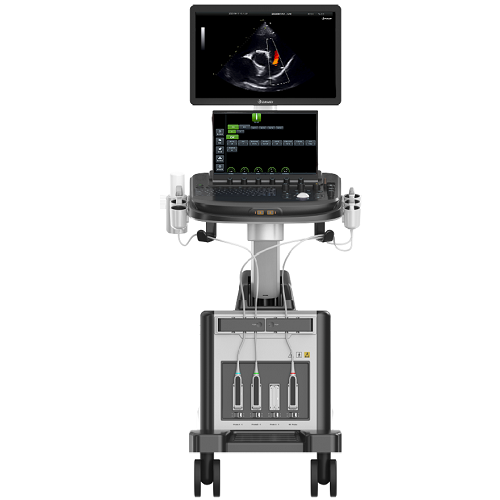
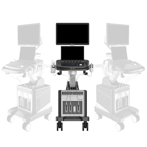
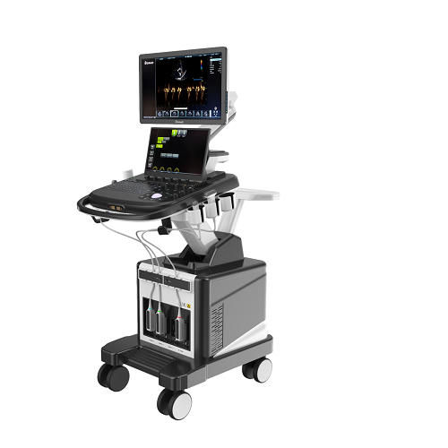
DWanimal T9-VET Veterinary Color Doppler Ultrasound
DWanimal T9-VET Veterinary Color Doppler Ultrasound
Couldn't load pickup availability
|
1.Product type: Veterinary Color Doppler Ultrasound Diagnostic System |
|
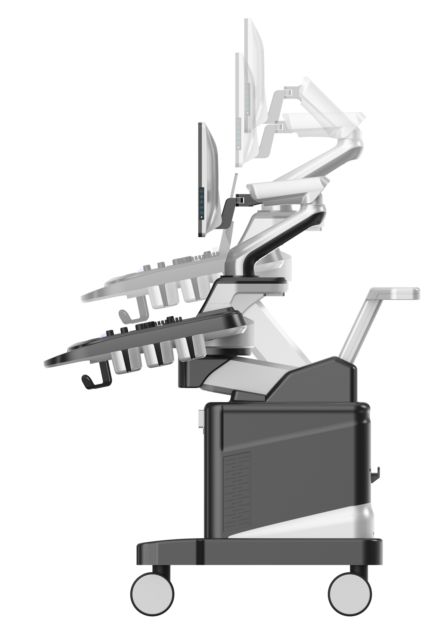
1.1Structural type: dual screens cart ultrasound |
|
2.Instructions for use and requirements: 2.1Meets the needs of pet hospitals, universities, colleges, and scientific research institutions for examination and diagnosis of the digestive system, reproductive system, urinary system, cardiovascular, musculoskeletal, ophthalmology, etc. |
|
3. Specifications and brief introductions |
|
3.1 Two-dimensional grayscale imaging 3.2 Color Doppler blood flow imaging 3.3 Spectral Doppler Imaging 3.4 M-type mode 3.5 B+C dual real-time mode 3.6 Tissue harmonic imaging 3.7 Spatial composite imaging technology 3.8 Real-time double-frame contrast imaging 3.9 Three simultaneous imaging (two-dimensional, color, spectrum simultaneous imaging) 3.10 Speckle noise suppression technology 3.11 Equipped with spatial composite imaging working mode 3.12 Main interface thumbnail 3.13 Full-screen Chinese and English annotation input, Chinese input method ≥ 2 3.14 Built-in case data management STATION workstation 3.15 Custom comments: including insertion, editing, saving, etc. 3.16 Canine body markings include 12 abdominal, 12 heart, and 9 genital markings; feline body markings include 12 abdominal, 12 heart, and 9 genital markings. 3.17 Operation interface: The host includes 2 operation language interfaces, Chinese and English. 3.18 comes standard with ≥21.5-inch high-resolution color LCD monitor 15.6-inch color LCD touch screen, supports multi-touch 3.19 The host has ≥4 built-in probe interfaces, all activated, consistent in size, and interoperable. 4D grayscale mode 4.1 Imaging modes include: B mode, M mode, supporting dual-window real-time display 4.2 Real-time image enlargement 4.3 Sound beamformer: digital sound beamformer, digital full-range dynamic focusing, digital dynamic variable aperture and dynamic apodization, and the focus position is fully adjustable in the imaging area 4.4 TGC segment adjustment 8 segments 4.5 pseudo color ≥8 kinds 4.6 TGC≥8 segments 4.7 Gain adjustment: B/M are independently adjustable, -45 dB~45dB, step 1 4.8 Dynamic range adjustment: 30dB~100dB, step 5 4.9 Maximum display depth ≥410mm 4.10 Partial magnification |
|
5 Color Doppler Mode: 5.1 Doppler gain continuously adjustable 5.2 It has the B+C function of simultaneous display of left and right sides on the same screen |
|
6. Spectral Doppler Mode 6.1 Transmission mode: pulse wave Doppler PW, continuous wave Doppler CW 6.2 PW test range 0~5m/s 6.3 CW test range 0~20m/s 6.4 Maximum measurement speed: forward or reverse blood flow speed 20m/s (CW) 6.5 SV sampling width and position range: width 1–8mm; 6.6 Display control: reverse display (left/right; up/down) |
|
7. Probe specifications 7.1 Probe configuration: linear probe, phased probe, micro-convex probe 7.2 Micro-convex probe: 3.0-11.0MHz 7.3 Linear probe: 3.0-12.0MHz 7.4 Cardiac probe: 3.0-10.0MHz 7.5 Cat-specific Cardiac probe: S12-4MHZ 7.6 Rat-specific linear array probe: 18MHZ 7.7 Linear probe: number of array elements ≧128 array elements; 7.8 Micro-convex probe: number of array elements ≧128 array elements; 7.9 Cardiac probe: number of array elements ≧ 64 array elements; |
|
8. Measurement/Analysis and Reporting: 8.1 Including depth, length, double length, trajectory circle, trajectory (closed), trajectory (non-closed), dual trajectory, angle, ellipse, standard circle, volume measurement, heart rate, speed, time, etc. 8.2 Obstetrics: Rich and dedicated canine and feline obstetrics measurement packages 8.3 Cardiology: Special cardiac measurement package, mitral valve, tricuspid valve, aorta, pulmonary vein, lung, Shunts score 8.4 Urinary System Package 8.5 Abdomen Measurement Software Package 8.6 Custom comments: including insertion, editing, saving, etc. 8.7 Standard case template |
|
9. Peripheral part: 9.1 Output interface: VIDEO interface, HDMI interface, RJ-45 interface, USB3.0 interface, S-VIDEO interface, DVI interface 9.2 Support network connection server 9.3 Support printers 9.4 Standard coupling agent heater |
|
10. Movie playback and raw data processing: 10.1 Movie playback, supports manual and automatic playback, you can select the playback duration (seconds) or the number of playback frames, and you can customize the duration or number of frames 10.2 The solid-state drive is 256G, which can permanently store videos and pictures, and can retrieve, transfer, and delete images at any time. 10.3 Multiple export image formats: dynamic images and static images can be directly exported in PC format, and the images can be viewed directly on an ordinary PC without special software. While exporting and backing up image data, real-time inspection can be performed without affecting the inspection operation. 10.4 supports DVD R/W burning optical drive 11.Standard configuration 11.1 Trolley-type cardiac ultrasound host 11.2 C3.0-11.0MHz Microconvex Probe 11.3 L3.0-12.0MHz linear array probe 11.4 P3.0-10.0MHz cardiac probe 11.5 Coupling agent heater 12. Optional accessories 12.1 ECG module with USB interface 12.2 Cat-specific heart probe: S12-4MHZ 12.3 Rat-specific linear array probe: 18MHZ 13. Product warranty: three years for the main unit and two years for the probe |
Share


